Innovative Approaches to Managing Cholesteatoma

Innovative Approaches to Managing Cholesteatoma

Innovative Approaches to Managing Cholesteatoma

Cholesteatoma, a destructive and expanding pathology within the middle ear and mastoid process, can precipitate grave complications, including the rare but potentially fatal temporal lobe abscess. The management of cholesteatoma, particularly in the context of an associated temporal lobe abscess, necessitates a meticulous, multidisciplinary approach, integrating surgical expertise with comprehensive medical management.

Recent years have witnessed significant advancements in surgical methodologies and diagnostic modalities, markedly improving patient outcomes in these complex scenarios. A notable case study by Garov et al. (2022) delineates the application of a combined extradural subtemporal and transmastoid approach in the successful resolution of intracranial complications stemming from otogenic origins. This innovative surgical strategy has proven effective in concurrently managing the temporal lobe abscess and the underlying cholesteatoma.

Early diagnosis and prompt intervention are paramount in addressing cholesteatoma-related temporal lobe abscesses. Baryshevskaya et al. (2020) underscore the imperative of an integrated approach that encompasses radiological imaging for accurate diagnosis and staging, followed by appropriate surgical intervention. This methodology not only facilitates the comprehensive removal of the cholesteatoma but also ensures the timely identification and management of the abscess.

Surgical Interventions

Surgical intervention typically focuses on two primary objectives: the eradication of the cholesteatoma and the drainage or excision of the abscess. C. Dai (2016) elaborates on the infratemporal fossa approach, which entails facial nerve rerouting for extensive cholesteatomas, allowing for complete removal of the pathology while preserving vital ear structures.

The intracranial extension of cholesteatoma, although rare, presents a significant clinical challenge. Waidyasekara et al. (2015) discuss a case of an extensive temporal lobe cholesteatoma, highlighting the criticality of early detection and aggressive surgical management. Managing such cases often necessitates a collaborative effort between otolaryngologists and neurosurgeons, emphasizing the need for a multidisciplinary approach.

Furthermore, the management of temporal bone fibrous dysplasia coexisting with external auditory canal cholesteatoma and temporal lobe abscess poses unique challenges, as illustrated by Zhang et al. (2020). These complex cases require tailored treatment strategies, potentially including canaloplasty for the external auditory canal cholesteatoma, followed by craniotomy for abscess drainage and resection of the dysplastic bone.

Postoperatove Care

Postoperative care is critical, with patients requiring vigilant monitoring for potential complications and recurrence. Antibiotic therapy is customized based on the culture and sensitivity patterns of the abscess, and regular follow-ups with imaging modalities like MRI or CT scans are essential to surveil for any recurrence of cholesteatoma or residual abscess.

Conclusion

In conclusion, the management of cholesteatoma in the setting of a temporal lobe abscess is a complex interplay of understanding the pathophysiology, possessing advanced surgical skills, and employing a holistic multidisciplinary approach. Prompt intervention, precision in surgical technique, and diligent postoperative care form the foundation of successful treatment outcomes. Ongoing research and the evolution of surgical techniques continue to enhance the prognosis for patients with this challenging condition, fostering optimism for improved outcomes and quality of life.

Innovative Approaches to Managing Cholesteatoma 2

Innovative Approaches to Managing Cholesteatoma Questions:

1. **Question:** A 45-year-old patient presents with a history of chronic otitis media and a recent diagnosis of temporal lobe abscess secondary to cholesteatoma. What is the first-line surgical approach for managing this condition?

A) Transmastoid approach
B) Endolymphatic sac surgery
C) Stapedectomy
D) Cochlear implantation
E) Tympanoplasty

**Answer:** A) Transmastoid approach
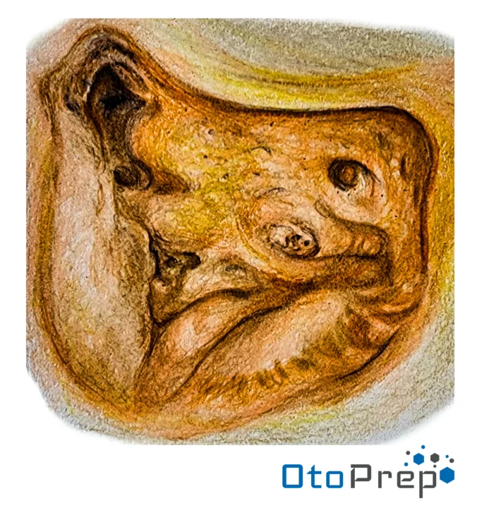
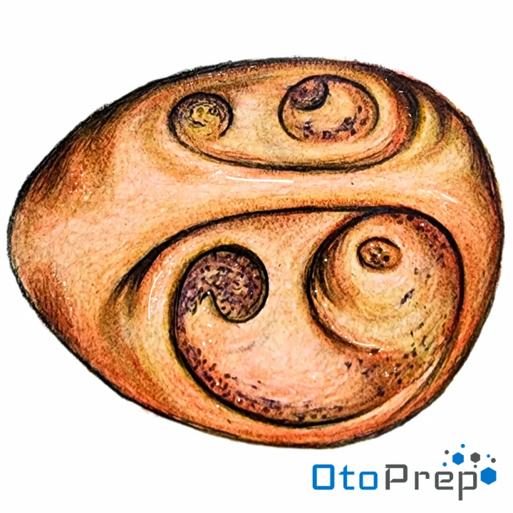
**Explanation:** The transmastoid approach is commonly used for surgical management of cholesteatoma, especially when there is intracranial involvement, such as a temporal lobe abscess.

2. **Question:** In the management of cholesteatoma-associated temporal lobe abscess, which imaging modality is most crucial for preoperative planning?

A) Plain X-rays
B) MRI
C) PET scan
D) CT scan
E) Ultrasound

**Answer:** D) CT scan
**Explanation:** A CT scan is essential for assessing the extent of cholesteatoma and its relationship to surrounding structures, crucial for surgical planning.

3. **Question:** A patient with cholesteatoma and temporal lobe abscess develops facial nerve palsy postoperatively. What is the most likely cause?

A) Anesthetic complication
B) Infection spread to the facial nerve
C) Iatrogenic injury during surgery
D) Postoperative hematoma
E) Reaction to antibiotics

**Answer:** C) Iatrogenic injury during surgery
**Explanation:** Iatrogenic injury to the facial nerve is a risk during surgery for cholesteatoma, particularly when the disease process involves the facial nerve canal.

4. **Question:** Which of the following is a potential complication of untreated cholesteatoma leading to temporal lobe abscess?

A) Sensorineural hearing loss
B) Meniere’s disease
C) Vestibular schwannoma
D) Meningitis
E) Tinnitus

**Answer:** D) Meningitis
**Explanation:** Untreated cholesteatoma, especially when complicated by a temporal lobe abscess, can lead to meningitis due to the spread of infection.

5. **Question:** Following successful surgical management of cholesteatoma with associated temporal lobe abscess, what is the most critical aspect of postoperative care?

A) Immediate cochlear implantation
B) High-dose corticosteroid therapy
C) Regular audiometric evaluations
D) Long-term antibiotic therapy
E) Vestibular rehabilitation exercises

**Answer:** D) Long-term antibiotic therapy
**Explanation:** Long-term antibiotic therapy is crucial to manage any residual infection and prevent recurrence, especially in cases where intracranial extension was noted.

European Board Examination in Otorhinolaryngology – Head and Neck Surgery (EBEORL-HNS)

Anatomy of the Neck for Otolaryngology Examinations

ebeorl-hns, european board of otolaryngology-head and neck surgery
European Board of Otorhinolaryngology Examinations: Professional Excellence in ENT

European Board of Otorhinolaryngology Examinations: A Gateway to Professional Excellence in ENT

The European Board of Otorhinolaryngology Examinations (EBEORL-HNS) represents a hallmark of quality and expertise in the field of ear, nose, and throat (ENT) care. Established by the UEMS ORL Section and Board in October 2008, EBEORL-HNS aims to harmonize the knowledge base among otolaryngologists in Europe, enhancing the free movement of specialists between member countries.

Overview of EBEORL-HNS

EBEORL-HNS operates as an independent entity, working in collaboration with various otolaryngology associations to develop and administer comprehensive examinations. These exams are designed to evaluate the skills, expertise, and competence of otolaryngologists, ensuring a high standard of care across Europe.

Certification Process

The certification process involves two parts: a written exam with 100 multiple-choice questions, and an oral exam. The written part must be passed to qualify for the oral exam. Successful candidates are awarded the title “Fellows of the European Board of ORL-HNS” or receive a “Diploma of the European Board of Otorhinolaryngology.”

Application and Exam Details for 2024

  • Registration: Open for the Part I online exam in 2024.
  • Exam Date: Part I is scheduled for Saturday, 20th April 2024.
  • Application Deadline: The deadline is 20th March 2024.
  • Exam Fee: 600€.
  • Eligibility: Candidates should be qualified specialists in ORL-HNS or in their final year of training.
  • Application Process: Candidates need to fill out an application form and provide all required official documents, translated into English.

Standardization and Training

Through its rigorous certification process, EBEORL-HNS ensures a standardized and high-quality level of training across Europe, contributing significantly to patient safety and care.

Collaboration and Professional Development

EBEORL-HNS maintains a close relationship with national otolaryngology societies, ensuring that its examinations reflect the latest advancements in the field. It also fosters continuous professional development among ENT specialists.

International Recognition

EBEORL-HNS certification is internationally recognized, facilitating professional mobility and exchange of expertise globally. It is an additional qualification to national education/exams, offering a range of benefits including proof of standard in UEMS affiliated countries, academic and job promotion, and higher salary prospects in some cases.

Conclusion

EBEORL-HNS plays a pivotal role in elevating otolaryngology standards in Europe. Its certification process, commitment to training standardization, continuous professional development emphasis, and international recognition mark it as a beacon of excellence in ENT care.

References

Keywords: EBEORL-HNS, European Board of Otorhinolaryngology, ENT Certification, Otorhinolaryngology Examinations, ENT Professional Excellence, Otolaryngology Training Europe, ENT Specialists Europe, Otorhinolaryngology Standardization, ENT Examination 2024, ENT Career Advancement, Otolaryngology Skills Assessment

Anatomy, Physiology, Pathology, and Pathophysiology of the Neck

Endolymphatic Sac Surgery for Otolaryngology Examinations

OtoPrep Endolymphatic sac surgical anatomy
Endolymphatic Sac Surgery Anatomy for Otolaryngology Examinations

Anatomy and Physiology

The endolymphatic sac is an extension of the membranous labyrinth, located in the posterior part of the temporal bone. It regulates endolymphatic fluid pressure and volume, crucial for inner ear function.

How well do you know Endolymphatic Sac Anatomy?

Identify these structures on the image above:
1. Horizontal semicircular canal
2. Facial nerve
3. Posterior semicircular canal
4. Endolymphatic sac
5. Posterior fossa dura
6. Sigmoid sinus
  • Horizontal semicircular canal: Detects angular acceleration.
  • Facial nerve: Responsible for facial expression and taste sensation.
  • Posterior semicircular canal: Also involved in detecting angular acceleration.
  • Endolymphatic sac: Regulates endolymphatic fluid pressure and volume.
  • Posterior fossa dura: Outermost layer of the meninges in the posterior fossa.
  • Sigmoid sinus: Drains blood from the brain towards the heart.

Pathology and Pathophysiology

Endolymphatic sac surgery is primarily indicated in the treatment of Meniere's disease, believed to be related to an imbalance in endolymph production and absorption.

  • Meniere's Disease: Characterized by vertigo, hearing loss, tinnitus, and aural fullness.
  • Endolymphatic Hydrops: Excessive accumulation of endolymph in the inner ear.

Endolymphatic sac surgery aims to reduce endolymphatic pressure through decompression or diversion of fluid. It may involve exposing the sac by removing bone and dura, followed by shunt placement or sac decompression.

Otolaryngology Examination Questions

  1. Indications for Surgery: "What conditions indicate endolymphatic sac surgery, and what are the expected outcomes?"
  2. Surgical Approach: "Describe the surgical approach to the endolymphatic sac for decompression in Meniere's disease."
  3. Complications: "What are the potential complications of endolymphatic sac surgery?"
  4. Anatomical Considerations: "Discuss the anatomical structures at risk during endolymphatic sac surgery."